From the PDF:
"Disease, if it is going to occur, develops 10 to 14 days after exposure. It is characterized by generalized hemorrhage, moderate to massive hepatic necrosis, and an immune-complex glomerulopathy. Characteristic karyomegalic changes and intranuclear inclusion bodies are typically found in macrophages and other antigen processing cells, including the mesangial cells of the glomerulus. The vast majority of birds with these lesions die. Adult birds and nestlings that are infected but do not develop disease will remain viremic for a variable period of time and shed virus in their droppings, and possibly in feather dander and skin, before becoming virus-negative. Most infected birds clear the virus after several weeks to several months and, although they maintain a persistent antibody titer, are not thought to be persistently infected"
Here is a chat GPT summary from this paragraph and others in the text:
"This disease appears in birds 10 to 14 days after exposure. It causes severe internal bleeding, liver damage, and kidney problems. Most birds with these symptoms do not survive. Some infected birds don’t get sick but can spread the virus through their droppings, feather dust, and skin for weeks or months before clearing the infection.
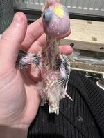
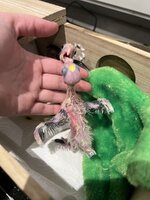
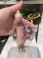
Different parrot species react differently to the virus. Conures younger than 6 weeks old and macaws and eclectus parrots under 14 weeks are the most at risk. Infected baby birds often seem healthy but suddenly die with little to no warning. Some signs before death include delayed digestion, weakness, pale or bruised skin, and, rarely, yellow urates. Blood tests, if done before death, may show liver damage and low platelet levels. Birds that die are usually in good body condition but may have an enlarged spleen and liver, internal bleeding, and fluid buildup in the abdomen or around the heart.
Some birds survive the initial disease but develop severe swelling (edema) and fluid buildup in their abdomen (ascites). Even though they remain alert, eat, and digest food, their condition does not improve. This may be due to low protein levels caused by liver failure or protein loss from the kidneys. These birds either die or must be euthanized. Unlike those that die suddenly, these birds do not have obvious viral inclusion bodies, making diagnosis harder. The disease closely resembles another viral infection (eastern equine encephalitis), which can cause confusion. However, these birds still carry a high amount of the virus, which can be detected in blood, cloacal swabs, or liver samples through PCR testing."

 And your little ones


